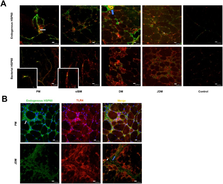
Figure 5. Interaction among HSP60, LC3 and TLR4 in IIM and control muscles.
(A) LC3-positive autophagosomes (red) are present in infiltrating cells (white arrow), within muscle fibers (asterisks), and blood vessels (blue arrows) (A, top panel). Vesicles positive for endogenous HSP60 (green) and LC3 (red) are present mainly in association with infiltrates and capillaries (A, top panel). Cells positive for bacterial HSP60 (green) in IIM muscles are also immunopositive for LC3 (red) (A, top panel). (B) Endogenous HSP60 (green) co-expresses with TLR4 (red) on some muscle fibers (asterisks), blood vessels (blue arrows) and in extracellular matrix (yellow arrows). Original magnification x40; bar: 20 µm. Original magnification of the insets x120; bars: 5 µm (PM) and 10 µm (sIBM).

