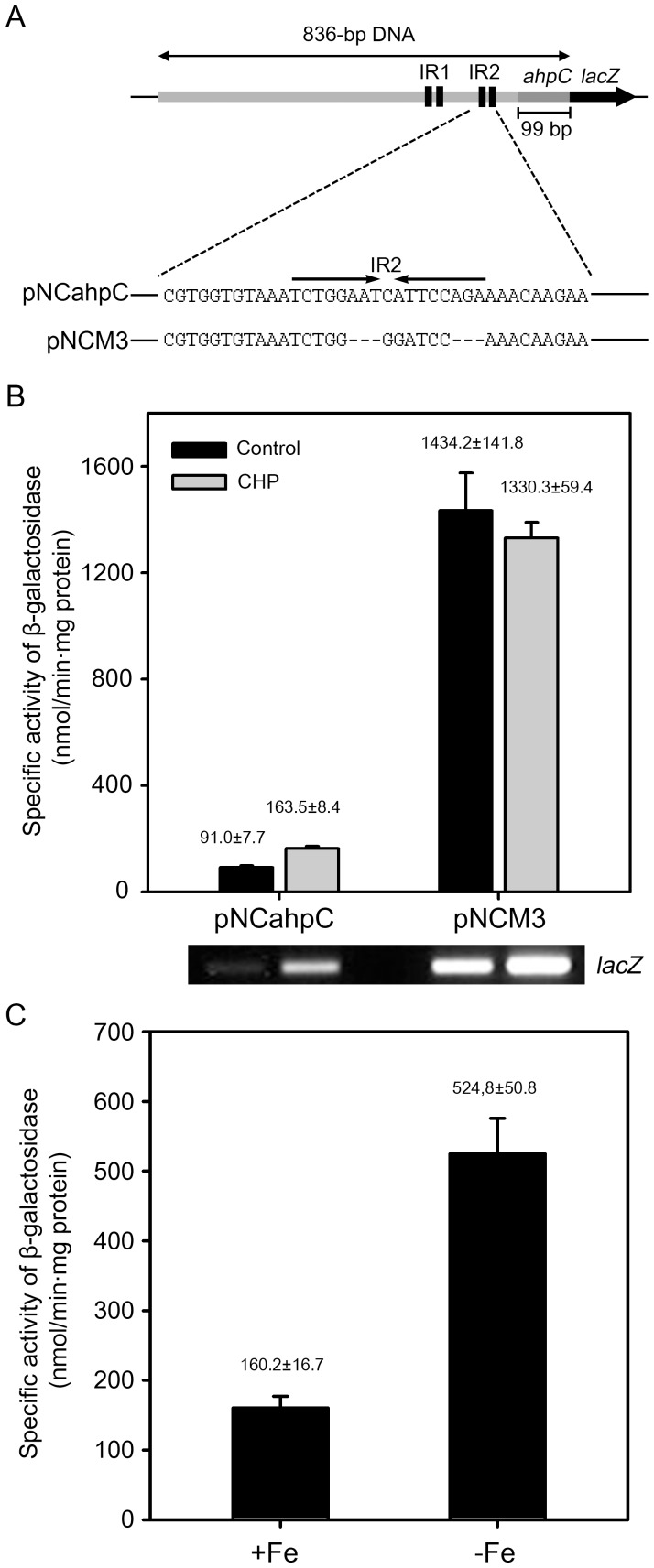
Figure 7. Effect of deletion of the IR2 sequence on ahpC expression and derepression of ahpC expression under iron-depleting conditions.
(A) Schematic diagram of pNCM3. The lacZ transcriptional fusion plasmid pNCM3 carries the same DNA fragment as pNCahpC except for the substitution of a part of IR2 with the BamHI recognition sequence. (B) M. smegmatis wild-type strains harboring pNCahpC and pNCM3 were grown to an OD600 of 0.45 to 0.5, and treated with CHP or DMSO (control). The cultures were further grown for 1 h. Cell-free crude extracts were used to measure β-galactosidase activity. Expression levels of lacZ in the wild-type strains carrying pNCahpC and pNCM3 were also determined by RT-PCR and the result is presented below the graph. All values are the means of two independent experiments. The error bars indicate the deviations from the means. (C) The wild-type strain of M. smegmatis harboring pNCahpC was grown in 7H9 medium to an OD600 of 1.5 to 2.0. Pre-cultured cells were washed twice with the original volume of MOPS medium supplemented with 0.02% Tween 80 and resuspended to the same volume of MOPS medium. 1 ml of the preculture was inoculated to 100 ml of MOPS medium supplemented with either 50 µM FeCl3 (+Fe) or 100 µM 2,2′-Dipyridyl (iron chelator) (−Fe). The strain was grown to an OD600 of 0.45 to 0.5 and harvested. Expression levels of ahpC were determined by performing β-galactosidase assay. The error bars indicate the deviations from the means of the two independent experiments.

