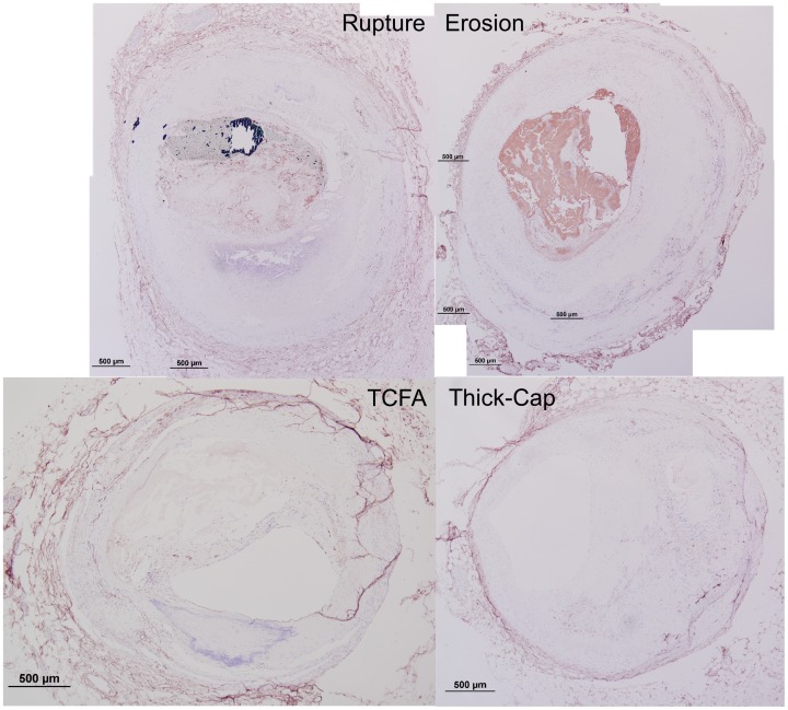
Figure 20. Representative histology images of CD31/CD34 staining.
Serial sections of the lesions shown in Figures 1 and 2 were stained for a combination of CD31 and CD34 (Nova Red chromagen) and counterstained with hematoxylin (blue) for nuclei. Artifacts such as the adventitial detachment in the TCFA specimen were manually masked out and excluded from the final analysis.

