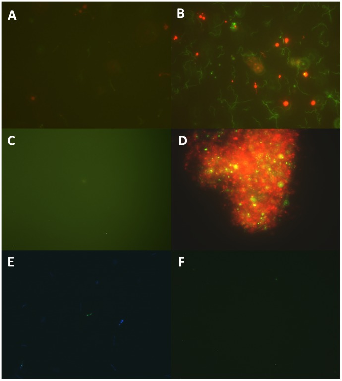
Figure 1. Representative images of B. burgdorferi culture (7 day old), observed with fluorescence microscopy equipped with Spot slider color camera using LIVE/DEAD BacLight stain (A), SYBR Green I/PI stain (B), and FDA stain (C).
Antibiotic-treated B. burgdorferi biofilm (9 day old) was stained by SYBR Green I/PI (D). Sytox Green/Hoechst 33342 stained B. burgdorferi images were recorded by the ORCA-R2 high resolution camera (E, merged from images visualized by DAPI, FITC and TRITC filters) and by Spot slider color camera with triple filter (F).

