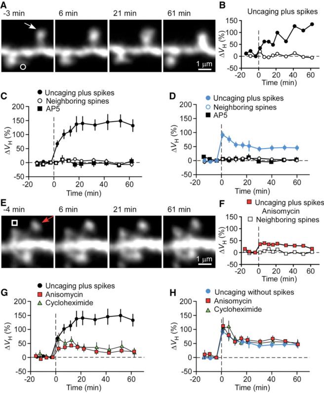
Fig. 1.
Spine-head enlargement induced by uncaging of glutamate with or without the application of postsynaptic spikes for single identified spines of CA1 pyramidal neurons in hippocampal slice culture. (A and E) Time-lapse z-integrated (z-stack) images of spines stimulated at time 0 by uncaging plus spikes in the absence (A) or presence (E) of anisomycin. Arrows indicate spots of two-photon uncaging of MNI-glutamate; open symbols indicate neighboring spines. (B and F) Time courses of changes in ΔVH for the stimulated (solid symbols) and neighboring (open symbols) spines shown in (A) and (E), respectively. (C and D) Averaged time courses of changes in ΔVH for spines stimulated by uncaging plus (C) or without (D) spikes in the absence (solid circles) or presence (solid squares) of AP5. Open circles represent data from neighboring spines in the absence of AP5 (open circles). Uncaging without spikes was performed in a Mg2+-free solution. Data are means ± SEM (n = 10 to 27 spines). The control trace shown in (C) and (G) was the average of 27 experiments performed in the same batches of slice preparations used for the test experiments. (G and H) Averaged time courses of changes in spine-head volume for spines stimulated by uncaging plus (G) or without (H) spikes in the absence (circles) or presence of anisomycin (red squares) or cycloheximide (green triangles). Data are means ± SEM (n = 7 to 27 spines).

