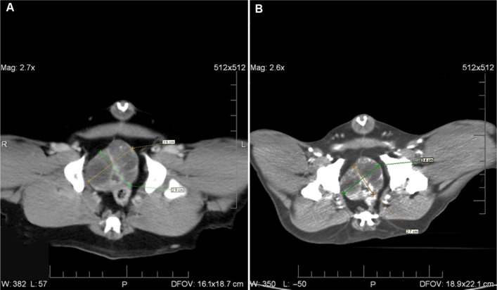
Figure 1.
Pre- and 4-week postinjection CT images of one of the dogs with prostatic carcinoma treated with GA-198AuNP that showed 50% tumor volume reduction.
Notes: (A) CT image of prostate carcinoma of the dog prior to treatment; (B) posttreatment CT image of same dog’s prostate tumor after 4 weeks. These images were chosen to represent the cross-section of the prostate at the same level; respiration, changes in bowel position, size and position of the urinary bladder, and the angle at which the pelvis is positioned affect the position of the prostate within the pelvic cavity between the two imaging studies. Scale bars representing 1 cm per division on these images are at the right hand side of the image. Prostatic volume was calculated in two different ways: the standard geometric formula for calculating the volume of an ellipsoid; and the length × width × height measurements times pi divided by 6 method commonly used in oncology. Measurements were also obtained from a three-dimensional computerized system which calculates the volume of the prostate based on the cross-sectional area and voxel thickness for each section of the prostate.
Abbreviations: CT, computed tomography; GA-198AuNP, gum arabic-coated radioactive gold nanoparticles.

