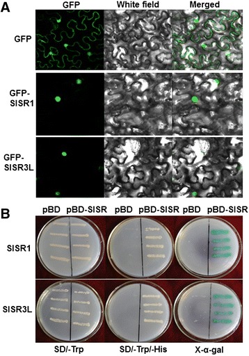
Figure 9.

Subcellular localization and transactivation activity of SlSR1 and SlSR3L proteins. (A) SlSR1 and SlSR3L are localized in nucleus. Agrobacteria carrying pFGC-Egfp-SlSR1, pFGC-Egfp-SlSR3L or pFGC-Egfp were infiltrated into N. benthamiana leaves and the images were taken in dark field for green fluorescence (left), in white field for the morphology of the cell (middle), and in combination (right), respectively. (B) SlSR1 and SlSR3L have transactivation activity. Yeast cells carrying pBD-SlSR1, pBD-SlSR3L or pBD empty vector (as a negative control) were streaked on SD/–Trp plates (left) or SD/–Trp/–His plates (middle) for 3 days at 28°C. The x-α-gal was added to the SD/-Trp/-His plates and kept at 28°C for 6 hr (right).
