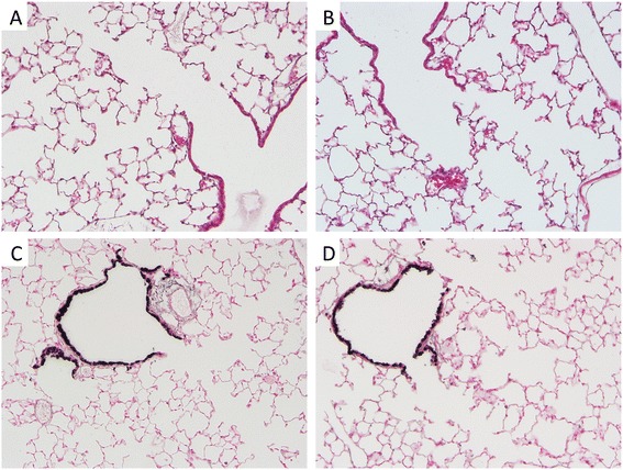
Figure 2.

RAGE TG mouse lungs had no significant histological alterations compared to control mouse lungs. Control lung (A) and RAGE TG sections (B) stained with H&E revealed no morphological disturbances. Immunostaining for CCSP, a marker of club (Clara) cells in the lung airway, revealed no qualitative differences when comparing normal control lung sections (C) with sections obtained from RAGE TG mice (D). Representative images (400× original magnification) of n = 3 mice in each group are shown.
