Figure 2.

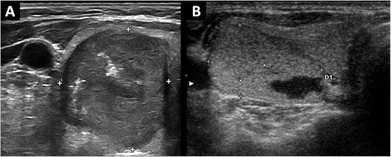
Different ultrasound presentations of MTC. (A) hypoechoic nodule with calcifications, classified at ultrasonography as suspicious. (B) mixed-spongiform nodule with hypoechoic halo, non-suspicious at ultrasonography.

Different ultrasound presentations of MTC. (A) hypoechoic nodule with calcifications, classified at ultrasonography as suspicious. (B) mixed-spongiform nodule with hypoechoic halo, non-suspicious at ultrasonography.