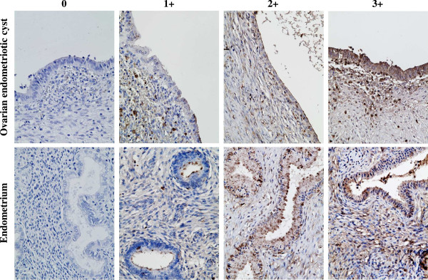
Figure 2.

Distribution of WFA-binding glycans in ovarian endometriotic cysts and normal proliferative phase endometrium. Both the glandular epithelial cells and the stromal component of ovarian endometriotic tissues as well as the normal proliferative phase endometrium were positive for WFA-binding glycans with strong to weak intensity depending on the samples (hematoxylin & eosin staining, original magnification ×40). Staining intensity for WFA-binding glycans assessed by histochemical staining score is summarized in Table 2.
