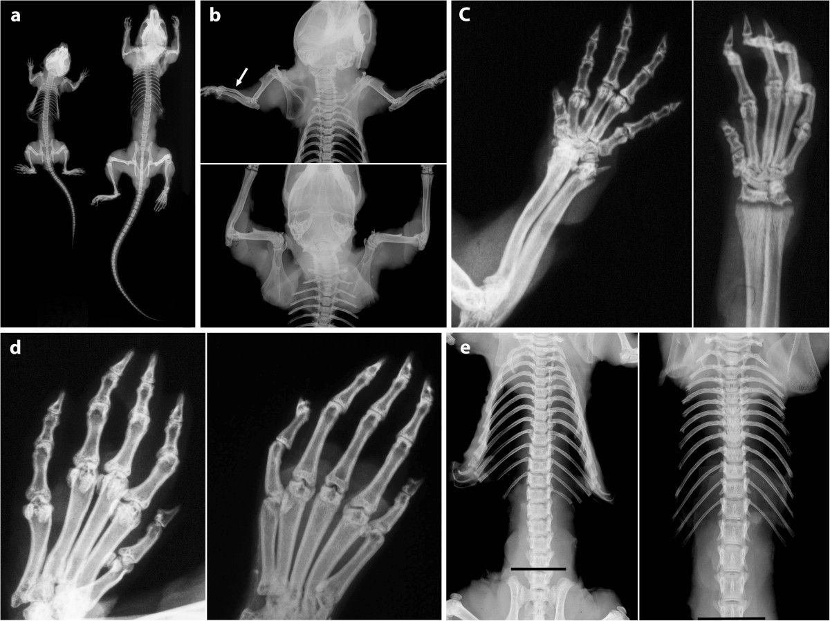
Figure 2.

Comparative Mouse Radiographs. Comparative radiographs of cn/+ and cn/cn mice at 5 weeks from the same litter are shown. Pairs of mice were radiographed together for size comparison. a: Entire skeleton is shown with cn/+ (right) and cn/cn (left). Other than being smaller, vertebrae (cervical to sacral); ribs; pelvis; hip, knee, and shoulder joints; and tail vertebrae are normal in cn/cn compared to cn/+ regarding numbers, shape, and structure. b: Radiographs show cn/cn (top) and cn/+ (bottom) forelimbs. Humeri of both are normal. Note curved mid-diaphyseal radius bilaterally of cn/cn (arrow) compared to straight shafts of radius and ulna of cn/+. Shortening of cn/cn long bones is evident. Note normal rib and thoracic vertebral segmentation and structure of both mice. c: Radiographs show radius and ulna and metacarpals and phalanges of cn/+ (right) and cn/cn (left). Distal radial and ulnar growth plates are clearly visualized on both views (b,c) from cn/+ but are not seen due to distal deformity on both views in cn/cn. Metacarpals and phalanges are shorter in the cn/cn but are not otherwise deformed compared with cn/+. d: In dorsoventral views of metatarsals and phalanges [cn/cn (left), cn/+ (right)], other than shortness of the cn/cn bones, basic shape is the same and there are no bulbous deformities of cn/cn metatarsals or phalanges. e: Specimen radiographs of ribs and spine illustrate cn/+ (right) and cn/cn (left) littermates. Rib and vertebral segmentation is normal in both with the shorter length characterizing cn/cn. Shorter cn/cn ribs cause a narrower ribcage and chest. Interpedicular widths in mice normally decrease from L1 to L6, unlike in the human where they progressively widen. Radiographs are shown from the same cervical vertebrae above. Lines below at same position in the 4th lumbar vertebrae demonstrate shorter stature of cn/cn mouse.
