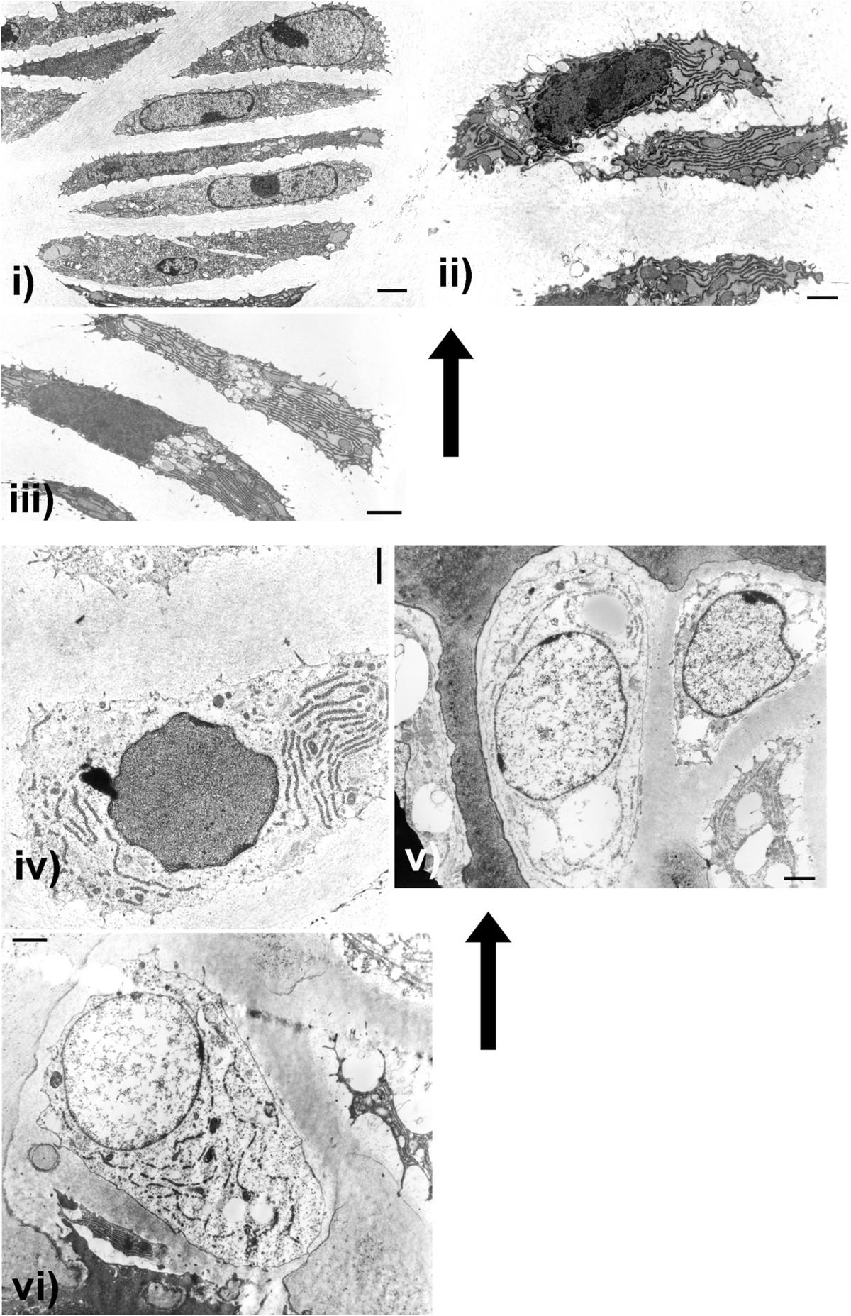
Figure 4.

Cn/cn Electron Microscopy. Electron micrographs (EMs) illustrate chondrocyte sequential changes from upper proliferating (palisading) zone (i) to lower hypertrophic zone (vi) in cn/cn physes. Cn/cn hypertrophic zones were markedly shortened compared to cn/+ but sequential individual cell changes throughout physes were similar. Arrows point to cells closer to upper regions (proliferating zone) of physes with lower regions (hypertrophic zone) below. EMs from cn/cn distal femoral and proximal tibial physes at 2.5 and 4.5 weeks are shown; proliferating zone (i to iii) and progressively lower cells in hypertrophic zone (iv to vi). Upper proliferating cell layers (i, ii and ii) show active flattened chondrocytes with abundant, well organized rough endoplasmic reticulum (RER) with dilated cisternae containing a homogeneous material indicative of active protein synthesis. In the cn/cn chondrocytes there were neither massively dilated collections of RER or abnormal electron dense collections within the RER which, if present, would indicate an ER storage disease. The hypertrophic cells in iv-vi show progressive hypertrophy, diminution of RER organelles but with some RER present even at the lowest levels at the metaphyseal junction. The hypertrophic cells in both sequences show well preserved organelles with most cells filling their lacunae with cell membranes intact. Line markers: i) 2.20 μm; ii) 1.00 μm; iii) 1.55 μm; iv) 1.26 μm; v) 1.55 μm and vi) 1.55 μm.
