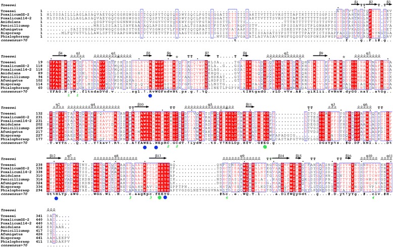
Figure 1.

Amino acid sequence alignment of endo-β-1, 4-mannosidase from P. oxalicum GZ-2 with related fungi. The secondary structural elements (α-helices displayed as squiggles, β-strands rendered as arrows, and strict β-turns as TT letters) of rPoMan5A are using Trichoderma reesei β-mannanase as a template (pdb no. 1QNO_A). The alignment includes endo-β-1, 4-mannanases from P. oxalicum GZ-2 (GenBank: AGW24296.1), Penicillium oxalicum 114-2 (GenBank: EPS31069.1), Penicillium sp. (GenBank: AFC38441.1), Bispora sp. MEY-1 (GenBank: ACH56965.1), Phialophora sp. (GenBank: ADF28533.1), Aspergillus fumigatus (GenBank: ACH58410.1), Aspergillus nidulans (GenBank: AGG69666.1), and Trichoderma reesei QM6a (GenBank: XP_006962944). Strictly conserved residues are highlighted with a red background and conservatively substituted residues are boxed. Pairs of Cys residues that formed disulfide bonds are indicated by green-colored numbers. The conserved catalytic residues Glu320 and Glu391 are indicated by a green dot and other conserved amino acid residues are marked with a blue dot. The figure was made using ESPript 3.0.
