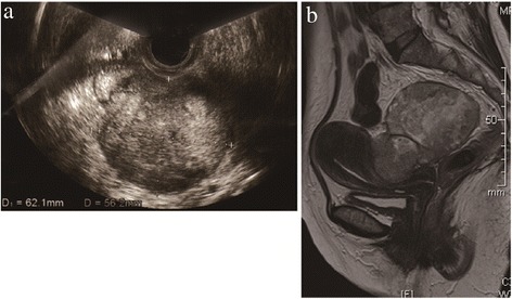
Figure 1.

Clinical imaging of the tumor. a. Vaginal ultrasonography revealed a 6 cm heterogeneous tumor located in the pouch of Douglas, with a suspected left ovarian involvement. b. T2-weighted MRI revealed an 8 cm heterogeneous tumor located at the posterior (intestinal) surface of the uterus, with a suspected rectal invasion.
