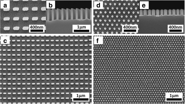
Figure 8.

SEM images of Si nanostructures generated by SRNIL and MCEE. (a,b,c) Close-up, cross-section, and overview of a 300-nm period square array of 190 ± 3 nm by 95 ± 2 nm rectangular cross-section Si nanopillars. (d,e,f) Corresponding views of a 150-nm period hexagonal array of sub-50-nm (46 ± 2 nm) diameter cylindrical Si nanopillars.
