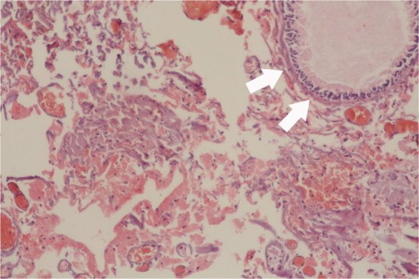
Figure 3.

Histopathologic microphotograph of the benign cyst wall. The microphotograph shows a benign cyst lined by ciliated columnar mucin-secreting cells (white arrows) with no secondary changes due to infection or hemorrhage; dyed with hematoxylin and eosin stain under 40 × magnification.
