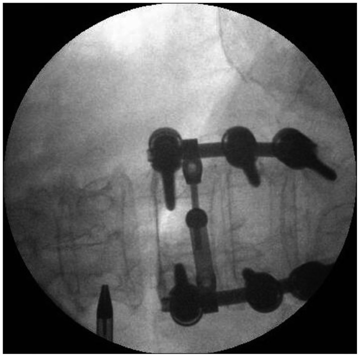
Fig. 5.
Intraprocedural fluoroscopic image showing cannula location during endoscopic RF denervation of L3 medial branch. First, 18 G needle was docked onto target point, at the junction of transverse process and superior articular process, and its position was confirmed on C-arm images. Then working cannula was inserted through the trajectory made by 18 G needle. RF : radiofrequency.

