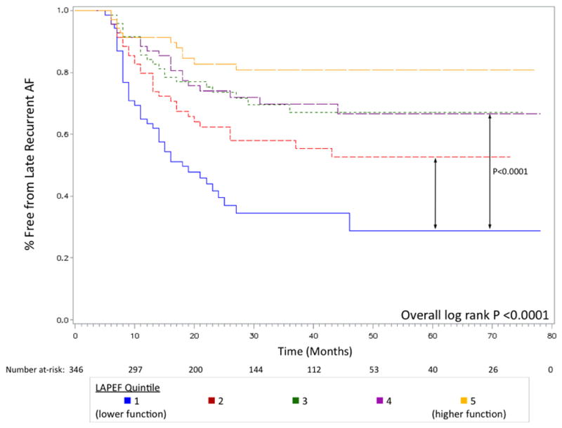
Figure 3. Kaplan-Meier survival curves for late recurrent AF after PVI stratified by LAPEF showing event-free survival time among five quintiles of function.

Patients with the highest LAPEF (“best function”, shown in orange) had the lowest recurrence rate over a median follow-up of 27 months. Selected P values for comparisons between groups are shown (quintile 1 vs. quintile 2, P=0.0113; quintile 1 vs. quintile 3, P<0.0001).
