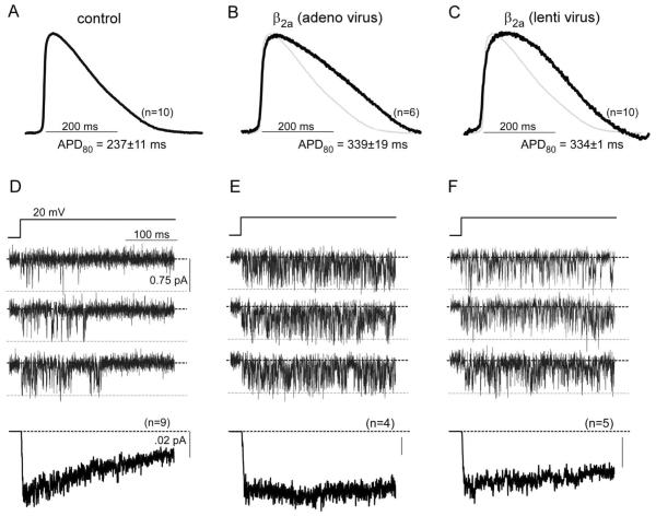
Fig. 3.
Genetic manipulability of aGPVM cultures by Ca2+ channel subunits. A: Control AP averaged across n = 10 syncytial aGPVM monolayers. Steady 2 Hz pacing. D: Corresponding single L-type Ca2+ channel activity from parallel cultures to those in A. Top, exemplar unitary Ba2+ currents evoked by +20-mV voltage step. Bottom, ensemble average current calculated from n = 9 such patches. Decay illustrates VDI. B, E: AP and Ca2+ channel activity following short-term expression of β2a-GFP construct via adenoviral transduction. Format as in A and D. Continued activity throughout voltage step indicates suppression of VDI. C, F: AP and Ca2+ channel activity following long-term expression of β2a-GFP construct via lentiviral transduction. Format as in A and D. Continued activity throughout voltage steps also indicates suppression of VDI.

