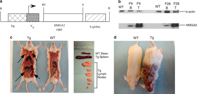
Figure 1.
(a) Eμ-HMGA2 construct. (b) HMGA2 expression in B and T cells of two transgenic lines. Western blot analysis was carried out using anti-HA antibodies. WT spleen lysate from a wild-type FVB/N mouse. F9 and F28 spleen lysates from F9 and F28 transgenic lines correspondingly. (c) Gross pathology of a representative 5-month-old Eμ-HMGA2 transgenic mouse, exhibiting greatly enlarged lymph nodes (arrows) and spleen relative to a wild-type of the same age. (d) A representative Eμ-HMGA2 transgenic mouse with severe skin lesion (right) and wild-type counterpart (left).

