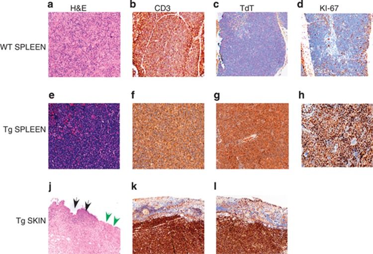
Figure 3.
Histopathological findings of adult Eμ-HMGA2 mice and wild-type control mice. The upper row demonstrates the spleen from a representative wild-type animal, using serial sections stained with hematoxylin and eosin (H&E) or labeled by immunohistochemistry (IHC) to reveal lymphocytes (the CD3 marker), TdT-positive cells and proliferating cells (Ki-67), whereas the middle row shows corresponding regions from the spleen of a representative HMGA2 transgenic mouse. The transgenic animal clearly exhibits a diffuse infiltration by myriad, proliferating neoplastic lymphocytes. The lower row shows the ulcerated skin from a transgenic mouse, indicating both ulcer (green arrows) and intact skin (black arrows). (a, e, j) Routine hematoxylin and eosin staining of WT splenic tissues, Tg splenic tissues and Tg skin lesions, respectively. (b, f, k) CD3 immunohistochemistry of WT splenic tissues, Tg splenic tissues and Tg skin lesions, respectively. (c, g, l) TdT staining of WT splenic tissues, Tg splenic tissues and Tg skin lesions, respectively. (d, h) KI-67 staining of WT splenic tissues and Tg splenic tissues, respectively.

