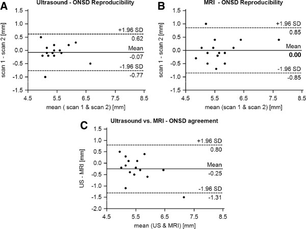
Figure 2.

Reproducibility and accuracy of ONSD assessment. Bland-Altman plots displaying the agreement of scan-rescan measurements of the optic nerve sheath diameter (ONSD) 3 mm behind the papilla by transorbital sonography (A) and MRI (B). Panel C demonstrates the agreement between sonographic and MRI-based ONSD quantification at the first visit. Continuous lines depict the mean of differences; dashed lines denote limits of agreement (mean ± 1.96 times of standard deviation).
