Figure 1.
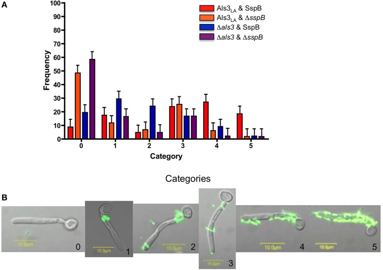
C. albicans and S. gordonii co-aggregation assays. (A) Experiments to assess co-aggregation between C. albicans and S. gordonii were conducted in a shaking flask as described (Silverman et al., 2010), with the exception that C. albicans cells from a 16 h yeast extract-peptone-dextrose culture were inoculated into RPMI 1640 medium for 90 min to form hyphae. These growth conditions were chosen because of our detailed knowledge of Als protein localization on cells cultured using this method (Coleman et al., 2009, 2010, 2012; Zhao et al., 2011). C. albicans strains included Als3LA (haploid for ALS3; encodes one wild-type copy of the ALS3 large allele from strain SC5314; Lin et al., 2014) and Δals3 (strain 1843, Δals3/Δals3 null mutant; Zhao et al., 2004). S. gordonii strains included SspB (wild type ATCC 10558) and ΔsspB (UB1360 Δ(sspA sspB); provided by Howard Jenkinson, University of Bristol; Silverman et al., 2010). Following incubation of C. albicans with S. gordonii, 100 C. albicans germ tubes were viewed microscopically and categorized to describe S. gordonii binding. The histogram shows the distribution of observations: category 0 (no bacterial cells attached to the germ tube); 1 (bacteria adhered only proximal to the mother yeast, a localization consistent with the involvement of Als1, Als2, and/or Als4); 2 (same as category 1, but with bacteria also at the germ tube tip, an alternate display for Als1); 3 (few bacteria bound diffusely across the germ tube); 4 (approximately half of the germ tube covered in bacteria); 5 (all or nearly all of the germ tube covered in bacteria). For each set of experiments, assays were repeated once or twice, on three or four different occasions. Data were analyzed using a mixed model analysis of variance. The mean for each category within a strain was analyzed using PROC MIXED in SAS (version 9.2, SAS Institute, Inc., Cary, NC). Separation of means was performed with the LSMEANS option. Differences were considered significant at P < 0.05. Means and standard errors are reported. The full set of comparisons between means is provided as Table 1 in Supplementary Material. (B) Fluorescent micrographs to illustrate the interaction between C. albicans and S. gordonii. C. albicans strain Als3LA was co-incubated with S. gordonii strain SspB as described above. Images are labeled with the number corresponding to the category in Figure 1A.

