Figure 4.
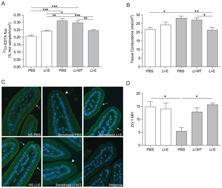
L. lactis expressing elafin(Ll-E) therapy protects NOD/DQ8 mice from gliadin-induced increases in paracellular permeability and preserves zonula occludens-1 (ZO-1) distribution. Sections of small intestine were mounted in Ussing chambers and (A) 51Cr-EDTA flux (% Hot Sample/h/cm2; n=8–11 per group) and (B) tissue conductance (mS/cm2; n=4–7 per group) was measured 24 h after the final gliadin challenge. (C) Sections of the proximal small intestine were stained for ZO-1 (green) expression. Nuclei labelled with 4′6-diamidino-2-phenylindole (DAPI; blue). Original magnification x20. White arrows indicate strong immunofluorescence at the apical junctional complex; arrowheads indicate patchy expression. (D) Mean fluorescence intensity (MFI) of ZO-1 staining in proximal small intestinal sections was determined (n=3–4 per group). MFI was corrected for background fluorescence. White bars represent non-sensitized animals, grey bars represent sensitized animals. Data are represented as mean ± SEM. *p<0.05, **p<0.01, ***p<0.001. Lactococcus lactis wild type (Ll-WT); L. lactis expressing elafin (Ll-E).

