
Fig. 1.
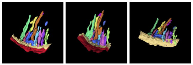
Three dimensional membrane structure of an avian cardiac muscle mitochondrion, obtained by electron tomography. The pleomorphic nature of the closely packed cristae is evident. The tilted view reveals the narrow openings of the cristae (crista junctions) into the boundary region of the inner membrane. The reconstructed sector of this mitochondrion is ca. 300 nm in length. Used with generous permission from Z. Almsherqi and Y. Deng.