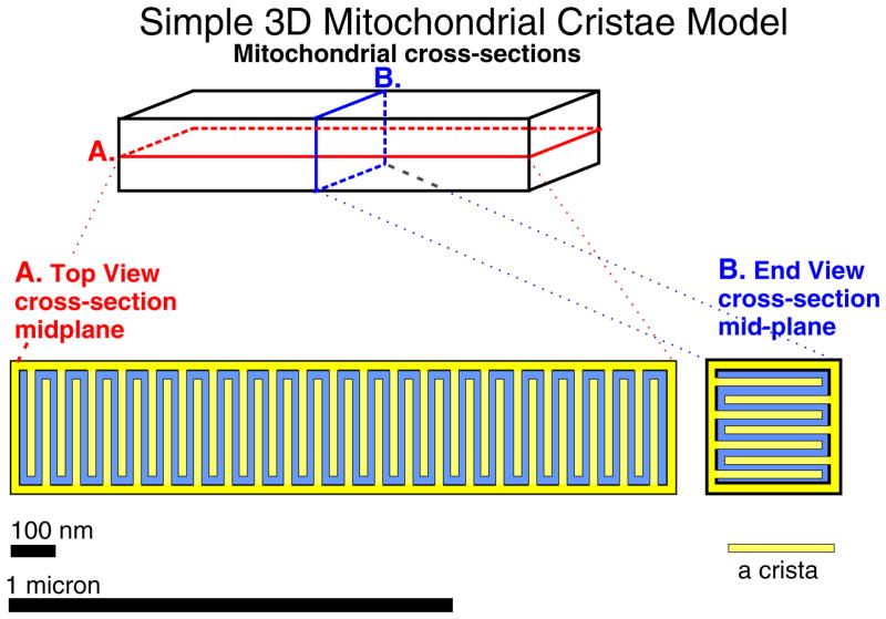
Fig. 4.
Simple mitochondrial cristae model in 3D. An intermyofibrillar mitochondrion (IFM) of size 1500 nm × 300 nm × 300 nm. A. Top view, cross-section, mid-plane. B. End view cross-section mid-plane. Each crista in the model is 240 nm long × 20 nm square and separated from other cristae by 20 nm. There are 42 cristae in a 40 nm plane and 7 layers of cristae, for a total of 294 cristae. The surface area per crista is 240 nm × 20 nm × 4 + 20 × 20 nm2 or 1.96 104 nm2. Thus the surface area of the inner membrane is 1.96 104 × 294 or 5.76 106 nm2, which is about three times larger than the surface area of the outer member, 1.98 106 nm2. The matrix is approximately half of the volume contained within the outer membrane. As noted, other shapes of the cristae may occur and will affect mitochondrial function.

