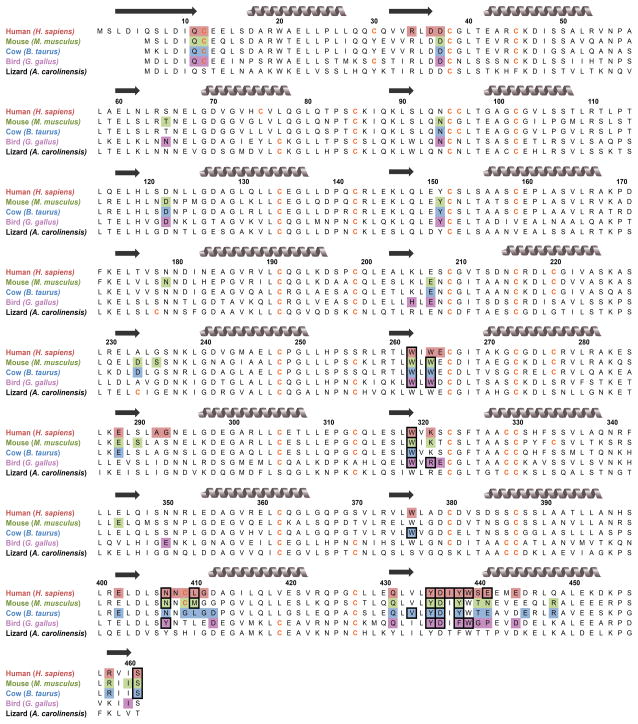
Fig. 4.
Amino-acid sequence alignment of homologous RIs. Residues participating in binding to endogenous RNases (as identified in crystal structures) are shaded. Black boxes indicate predicted “hotspots” for binding affinity [32]. Gray coils represent α-helices, and black arrows represent β-sheets.

