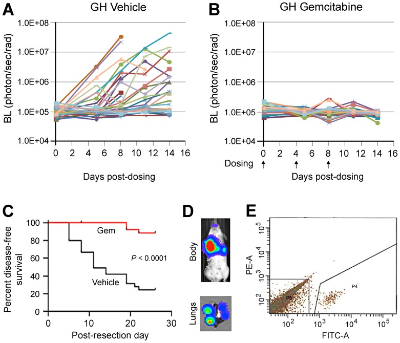
Figure 6. The GH mouse allows for consistent tracking of the progression of labeled metastases, their therapeutic responses and isolation in a preclinical adjuvant study.
ffLuc-eGFP-labeled LLC tumors were transplanted subcutaneously into GH mice. Upon reaching 500 mm3, the primary tumors were resected. A and B, GH mice were randomized into control and treated groups to receive vehicle and gemcitabine at 25 mg/kg, respectively. Metastatic progression in mice was periodically monitored by BL imaging. Metastatic growth was efficient in untreated GH mice (A) but suppressed by gemcitabine (B). C, Kaplan-Meyer analysis showed that survival was significantly shorter in control compared to treated GH mice. D and E, GFP+ cancer cells can be readily isolated from whole lungs of GH mice. The lungs were harvested from control GH mice, made into a single-cell suspension, and subjected to sorting by FACS to isolate GFP+ cells. The representative result from mouse #806 is shown. The in vivo image of mice and ex vivo image of lung (D) showed BL signal from pulmonary metastases and individual lung nodules, respectively. The GFP+ cancer cells were successfully isolated from whole lung by FACS sorting (P4 subpopulation in E).

