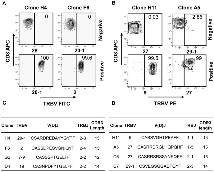
Figure 6. TRBV analysis of LCL- and tumour-reactive CD8+ T cell clones.
CD8+ T cell clones specific for (A, C) LCL or (B, D) tumour antigens were assessed for clonality by flow cytometric and molecular analysis. (A, B) Clones were stained with a panel of fluorescently-labelled TRBV mAb, then analysed on a flow cytometer. Positive and negative TRBV staining is shown for representative T cell clones. (C, D) To confirm the flow cytometric findings and determine unidentified TRBV regions, RNA was extracted from T cell clones and TRBV regions reverse transcribed, amplified, then sequenced. The designations TRBV and TRBJ follow the TCR gene nomenclature specified by IMGT [18].

