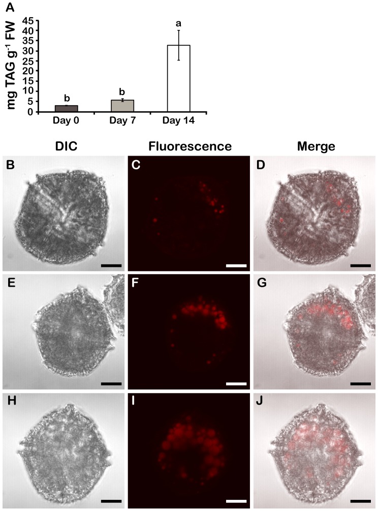
Figure 6. TAGs accumulate in N stressed cells.
A) Neutral lipid levels. Results are mean ± SE (n = 3). Statistically different results (p<0.05) are marked with a different letter (Analysis of variance). FW; Fresh weight. DIC, Nile red-stained lipid bodies and merged images of day 0 (B–D), day 7 (E–G) and day 14 cells (H–J). All cells were pictured from a ventral view. Lipid bodies were most predominant in the anterior part of the cells.

