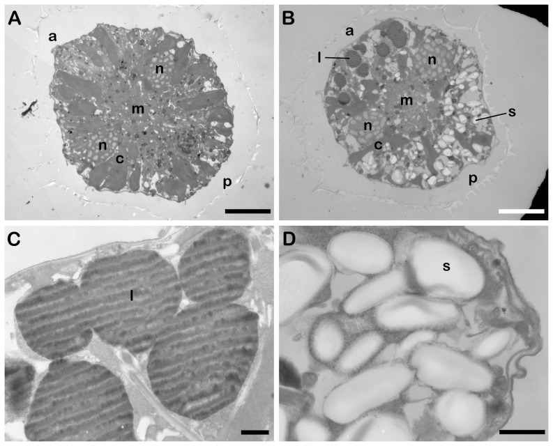
Figure 7. Polarized localization of lipid bodies and starch granules visualized by transmission electron microscopy.
Cross-sections of a cell at day 0 (A) and at day 14 in f/2-N medium (B). Both are in a ventral orientation and scale bars are 10 µm. Lipid bodies (l) are located predominantly at the anterior (a) end of the cell, while starch granules (s) are localized at the posterior (p) end. The ends of the C-shaped nucleus (n) surround a central Golgi/ER membrane region (m). Chloroplasts (c) are less abundant in day 14 cells. Higher magnification images of lipid bodies (C) and starch granules (D) in a cell at day 14. Scale bars are 1 µm.

