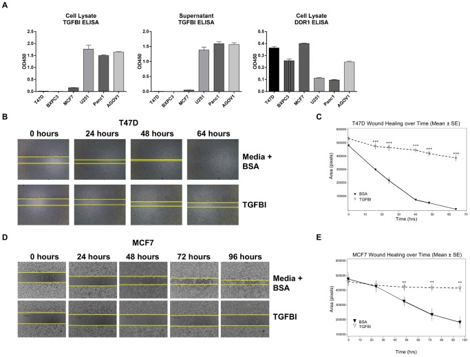
Figure 6. TGFBI inhibits lateral migration of T47D and MCF7 cells.
(a) A small panel of tumor cells was tested for TGFBI and DDR1 expression via ELISA. TGFBI ELISA was run with both cell lysates and supernatants; DDR1 ELISA used only cell lysates. Exogenous addition of TGFBI completely abolished wound healing capability of T47D (b) and MCF7 (d) as earlier observed with BXPC3. The area of wound closure was photographed over time. The area closed at different time points was measured using the Image J program. The wound healing over time is plotted for T47D (c) and MCF7 (e). In all cases, media with BSA was used as a negative control. BSA (▾) TGFBI (▽) The data are representative of three independent experiments, each run in triplicate. Statistical significance is reported as *** (p<0.0001) and ** (p≤0.005).

