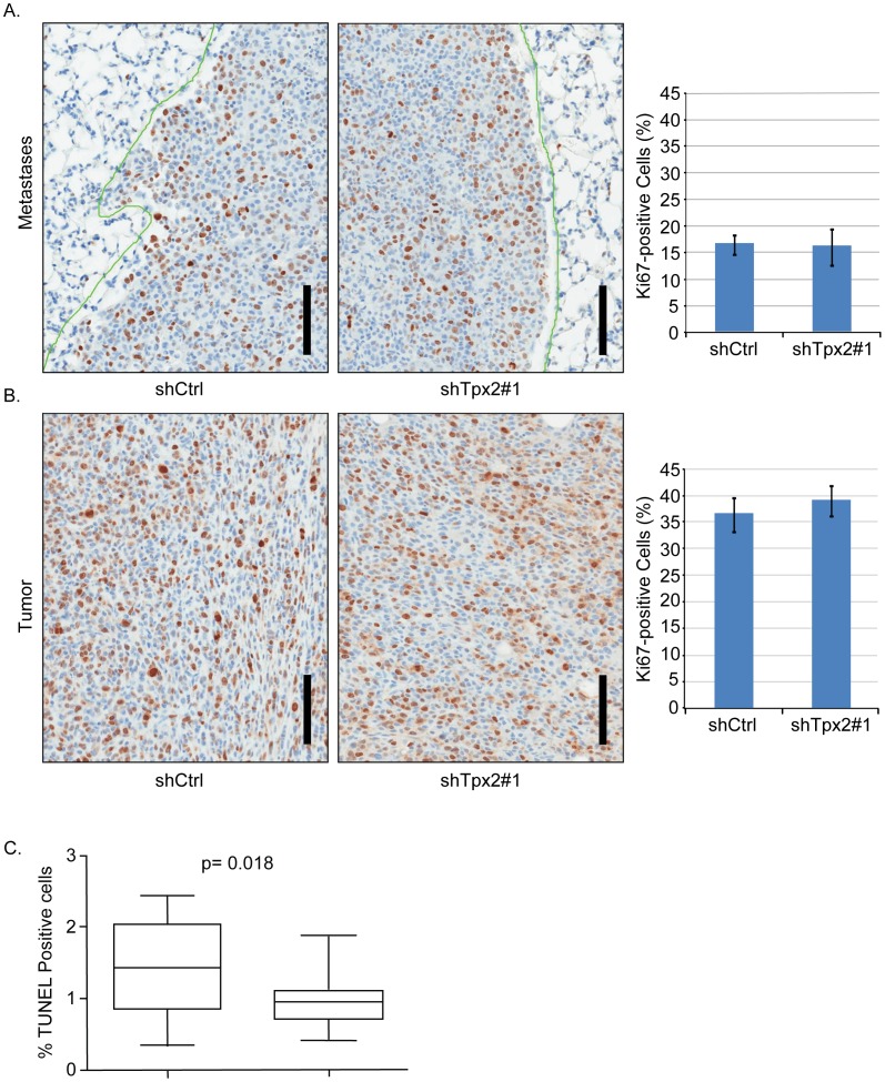
Figure 5. Knockdown of Tpx2 does not impair 6DT1 cell proliferation in vivo and does not affect anoikis.
A) Lung sections from mice injected with 6DT1-shCtrl or 6DT1-shTpx2#1 harboring metastatic nodules and immuno-labeled with Ki-67 antibody. The bar diagram on the right represents quantification of percent tumor cell nuclei with immuno labeling (cycling fraction) relative to total numbers of tumor cells from five mice each. Error bars show standard deviations. B) Sections of primary tumors from mice injected with 6DT1-shRNA control or 6DT1-shTpx2#1 were stained and analyzed as in a). Scale bars correspond to 100 µm. C) shCtrl or Tpx2 knockdown cells were plated into low adhesion plates, grown for 7 days, and viable cells quantified. The combined results of four independent experiments are represented. No significant difference in anoikis was observed by reduced Tpx2 levels.

