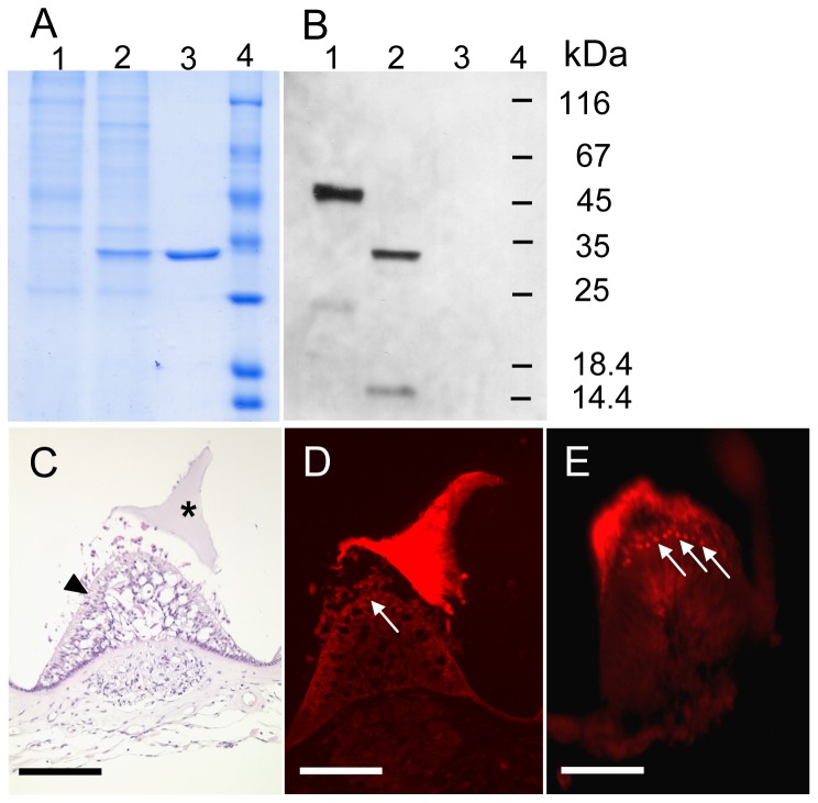
Figure 4. Immunodetection of zona pellucida-like domain protein from salmon samples.
A, Coomassie stained SDS-polyacrylamide gel and B, corresponding Western blot of separated crude cupula extracts. Lane 1, untreated sample, lane 2, PNGase F treated sample with faster migration of zona pellucida-like domain protein. C, HE stained inner ear cross section, asterisk marks the cupula and the arrowhead the subcupulary region with sensory and supporting cells. D, immunostaining of inner ear cross section, the arrow probably indicates staining of supporting cells which produce the zona pellucida-like domain protein. This is even more pronounced in E.

