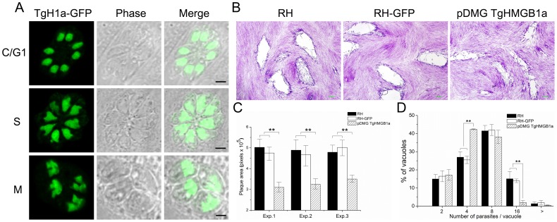
Figure 6. Overexpression of TgHMGB1a affects intracellular parasite replication.
A. Transgenic TgHMGB1a localized to the nucleus, similar to endogenous TgHMGB1a. Plasmids of pDMG TgHMGB1a (Figure S9A) were introduced into the RH strain to overexpress TgHMGB1a. Through screening, clones were obtained in which the TgHMGB1a-GFP was expressed and concentrated in the nucleus throughout the entire cell cycle (cytokinesis, C; gap phase, G1; DNA synthesis, S; and mitosis, M), which was identical to endogenous TgHMGB1a, as shown by IFA ( Figure 2E ). Western blot were applied to confirm the overexpression of TgHMGB1a (Figure S9B). B. Plaque assays for TgHMGB1a overexpression parasites compared to its parental strains. Infected HFF-monolayers were fixed and stained (see Materials and methods). Scale bar, 0.2 mm. C. Plaque assays were carried out three times independently, the plaque area was quantified from three independent experiments. At least 30 plaques were quantified per strain in every experiment. Each bar indicates the mean ± SD; “exp.” indicated the each experiment. D. An intracellular growth rate assay was carried out through counting the number of parasites per vacuole. Parasites of the TgHMGB1a overexpressed and parental strains (RH and RH-GFP) were allowed to invade and divide for 24 hours. IFA was performed with TgSAG1 abs and the numbers of parasites per vacuole were counted. At least 150 vacuoles were examined per strain. Each bar indicates the mean ± SD from three independent experiments; n≥150 vacuoles. Statistical significance was determined using Student's t-test (**P<0.05).

