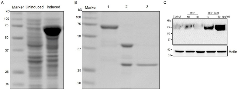
Figure 2. Expression, purification and characterization of MBP-TcpF.
(A) Cell lysates from uninduced and induced cultures of C41 (DE3) E. coli harboring plasmid pOU1811 were separated on a 10% SDS-PAGE gel and stained with Coomassie blue R-250. (B) Purified MBP-TcpF (lane 1), MBP-TcpF digested with enterokinase (lane 2) and purified TcpF (lane 3) were run on a 10% SDS-PAGE gel and stained with Coomassie blue R-250. (C) RAW264.7 cells incubated with increasing concentrations of MBP-TcpF or MBP alone for 5 h. After washing and treatment with trypsin, the lysate was subjected to Western blot and probed with antibodies to TcpF.

