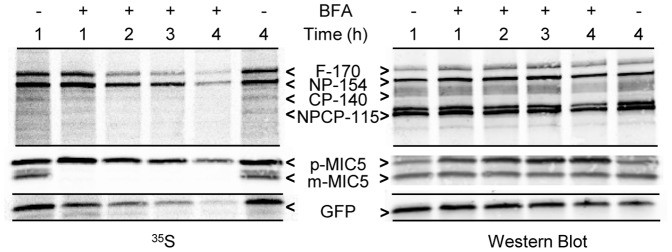
Figure 4. Protein synthesis during BFA treatment assessed by biosynthetic labeling of FtsH1, MIC5 and cytosolic GFP.

Fibroblast monolayers infected with T. gondii expressing FtsH1 tagged internally with V5 epitopes and a cytosolic GFP (∼108) were pre-incubated with or without BFA (1 µg/ml) for the indicated times prior to being labeled with 35S-methionine/cysteine for 30 minutes. Samples were immunoprecipitated with anti-V5 mAb, anti-GFP, and anti-MIC5 before being separated on 7.5% (FtsH1) or 8–16% (GFP and MIC5) SDS-PAGE gels and transferred to nitrocellulose. The left panel shows phosphorimaging, the right panel shows the same lanes detected by Western blot. The four major forms of FtsH1 are marked according to their apparent molecular mass on SDS-PAGE: full-length (F-170), N-terminally processed (NP-154), C-terminally processed (CP-140) or dual processed (NPCP-115). In a 30 min labeling, the first two forms predominate [21]. The precursor (p) and mature (m) forms of MIC5 [75] are marked.
