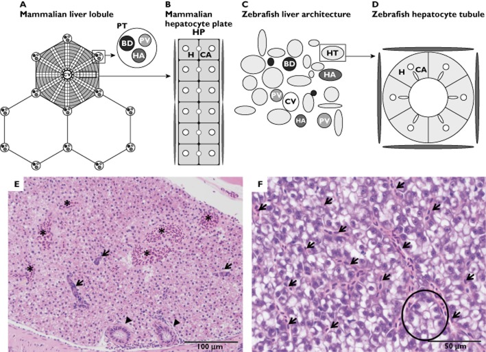
Figure 1.
Schematic transverse representations of mammalian and zebrafish liver architecture. (A) The mammalian liver lobule, arranged with plates of hepatocytes radiating outward from a central vein (CV). At the corners of each lobule are portal tracts (PT), containing a portal vein (PV), a hepatic artery (HA) and a bile duct (BD). (B) Mammalian bilayered hepatocyte plate. Bicellular canaliculi (CA) are located adjacent to the hepatocytes (H) in the hepatocyte plate (HP). A basal hepatocyte membrane allows transport of oxygen, proteins and different macromolecules to the hepatocytes. Blood enters the liver through the portal vein and hepatic artery, after which it enters the central vein through sinusoid vessels, located between the plates. (C) The zebrafish liver architecture. The portal vein (PV), hepatic artery (HA), bile duct (BD), hepatocyte tubule (HT) and the central vein (CV) are scattered throughout the parenchyma. (D) Zebrafish hepatocytes (H) are arranged in tubules around small bile ducts, which receive bile from the hepatocyte canaliculi (CA). Sinusoids are located at the periphery of these tubules. (E) Histological image of male zebrafish liver (haematoxylin and eosin staining at ×200 magnification). Note the presence of several biliary ducts (arrows), bile ductules (arrowheads) and blood vessels (*), with lack of lobular arrangement. (F) Histological image of female zebrafish liver (haematoxylin and eosin staining at ×400 magnification). This high-power image displays sinusoidal spaces between hepatocytes (arrows) and an instance of the tubular arrangement of hepatocytes (encircled), which is frequently not visible histologically. Note the difference in staining of male and female zebrafish liver

