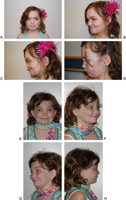
Fig. 3.

A 12-year-old girl with Crouzon syndrome who underwent a subcranial Le Fort III with distraction osteogenesis. (A) Preoperative photograph, anteroposterior (AP) view. (B) Preoperative photograph, left three-quarter view. (C) Preoperative photograph, left lateral view. (D) Early postoperative photograph, with the internal distractor in place, left lateral view. (E) Postoperative photograph, AP view. (F) Postoperative photograph, right three-quarter view. (G) Postoperative photograph, left three-quarter view. (H) Postoperative photograph, right lateral view.
