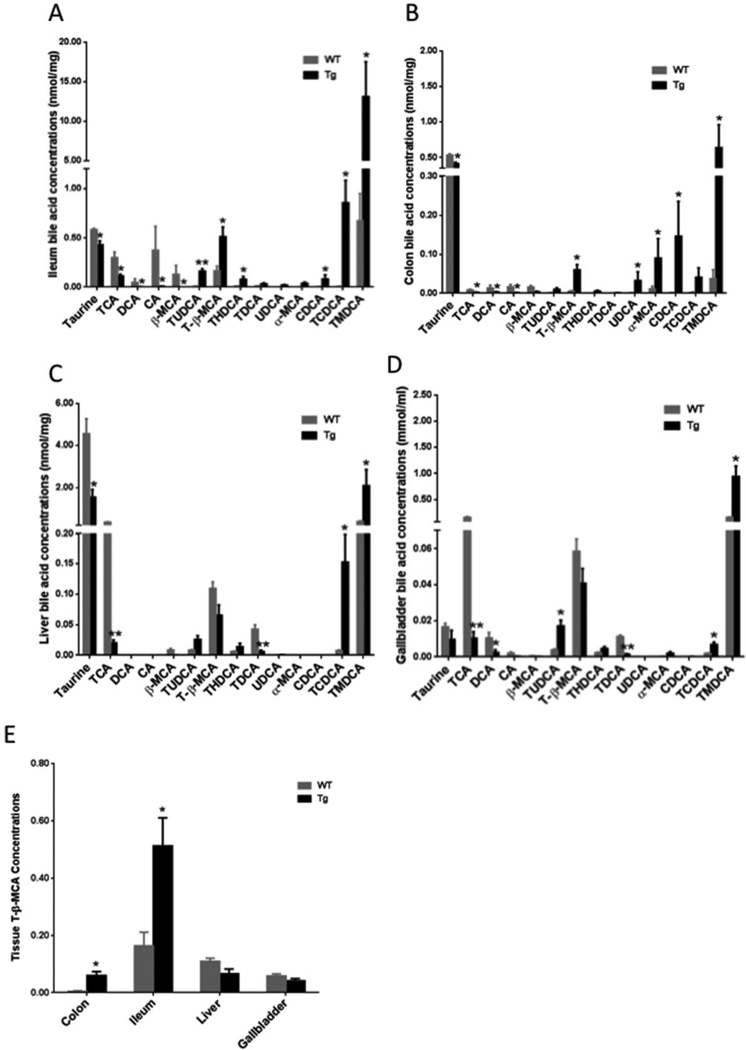
Fig 6. Quantitation analysis of the bile acid markers.
Bile acid marker levels in the ileum (A), colon (B), liver (C), and gallbladder (D) of female CYP7A1-tg and wild-type (WT) mice. (E) T-β-MCA levels in colon, ileum, liver and gallbladder samples (nmol/mg tissue for colon, ileum and liver, and mmol/ml for gallbladder). Data were expressed as mean ± SD. Significant comparison was based on two tailed Student’s t-test or Mann-Whitney test. An * indicates p < 0.05, and a ** indicates p<0.01 (with respect to the WT group).

