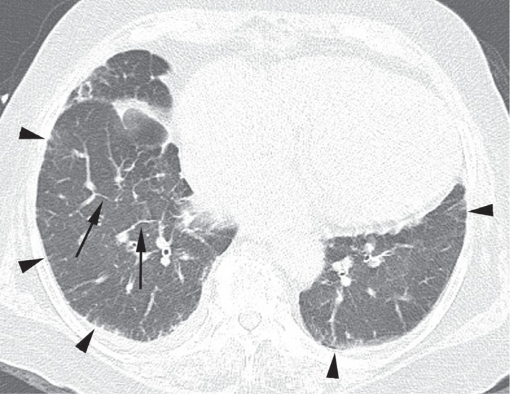
Figure 1.
A 74-year-old female with amiodarone pulmonary toxicity (APT) exhibiting a pulmonary interstitial fibrosis pattern. Computed tomography (CT) scans obtained at the level of both lower lobes revealed intralobular and interlobular septal thickenings in the peripheral regions of both lower lobes (arrowheads), and interlobular septal thickenings in the central and middle regions of the right lower lobe (arrows). The APT CT score was 4 on this CT section; the involved regions included the central, middle, and peripheral regions.

