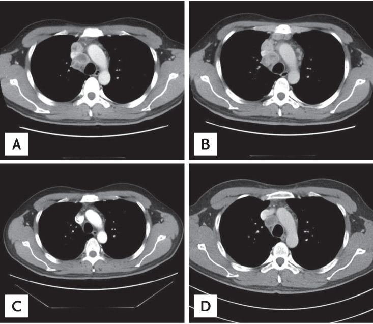
Figure 3.
Chest computed tomography images of paratracheal lymph nodes. (A) Paratracheal lymph node enlarged at the time of initial diagnosis. (B) Disease progression after two cycles of pemetrexed-cisplatin as first-line chemotherapy. (C) Partial response after 2 months of erlotinib. (D) Image before rebiopsy, showing the paratracheal lymph node had increased in size despite erlotinib treatment.

