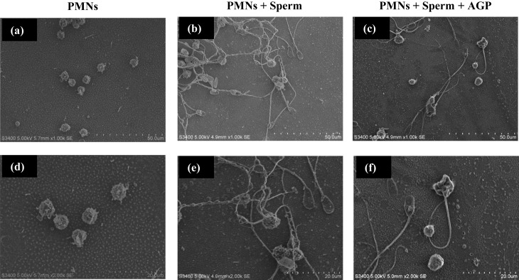
Fig. 2.
Scanning electron microscopy of sperm phagocytosis by PMNs. The upper panels are images obtained at ×1,000 (a, b, c) and the lower panels are those obtained at ×2,000, respectively. PMNs were incubated without any stimulant (a, d), or with sperm addition to induce neutrophil extracellular traps (NETs) (b, e). NET formation was suppressed in PMNs incubated with AGP (100 ng/ml) prior to a phagocytosis assay (e, f).

