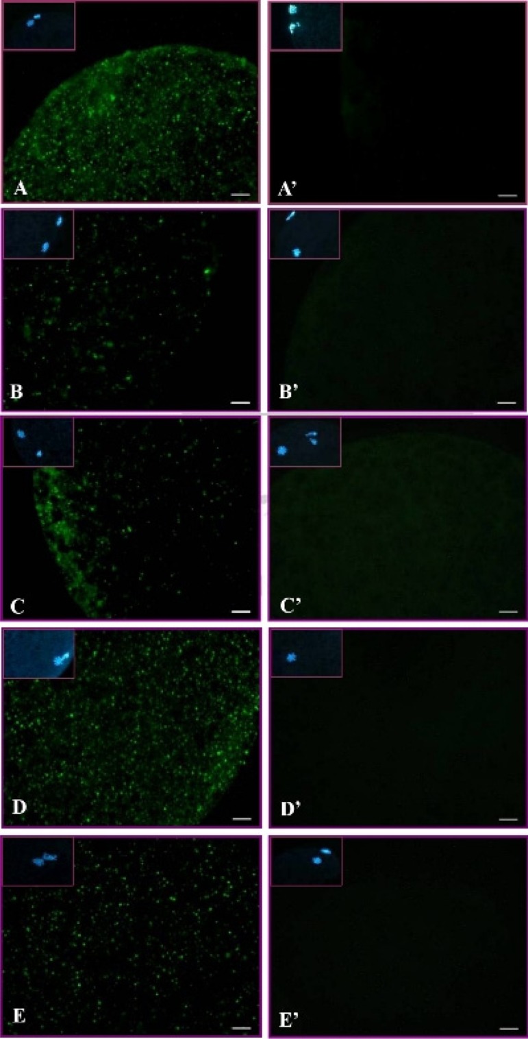
Fig. 4.

Subcellular localization of p-p38 in matured porcine oocytes in the non-heat-shocked (39 C) and heat-shocked (41.5 C) groups. Green dots are p-p38 labeled by immunocytochemical staining. (A–E) Expressions of p-p38 in matured oocytes after heat shock for 0 h (A), 1 h (B), 2 h (C) or 4 h (D) and the p-p38 level in matured oocytes cultured at 39 C for 4 h (E). Panels A’–E’ are negative controls (without primary antibody), and insets are chromatin/chromosome (blue) structures stained by Hoechst 33342. Scale bar, 10 μm.
