Figure 6.
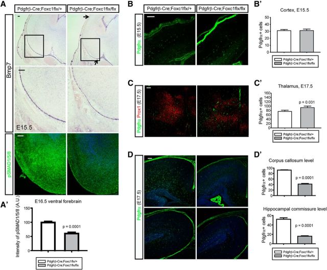
Meninges-mediated migration of OPCs into the cortex. A, To obtain the reduced meningeal expression of Bmps, a Pdgfrβ-Cre mouse was bred to Foxc1 conditional mutant to get Pdgfrβ-Cre;Foxc1flx/flx mutants. In situ hybridization of Bmp7 shows reduced Bmp7 expression in the meninges of ventral forebrain at E15.5 (arrows). Higher magnification images of boxed areas are presented in the middle. Staining of pSMAD1/5/8 shows the reduced activation of Bmp signaling in the mutant ventral forebrain at E16.5 (bottom). A′, A graph shows the intensity of pSMAD1/5/8 in the ventral forebrain (n = 4). A measure function of ImageJ software was used. B, B′, Cortical OPCs were stained for Pdgfrα and counted at E15.5. C, C′, Ventral OPCs were stained for Pdgfrα and counted at E17.5. Prox1 was used to stain the thalamic neurons to match the sections. p = 0.031 (Mann–Whitney rank sum test; n = 4). D, D′, Cortical OPCs were counted in the anterior (top) to the posterior (bottom) cortex (n = 4). Error bars depict SEM. Scale bars: 100 μm.

