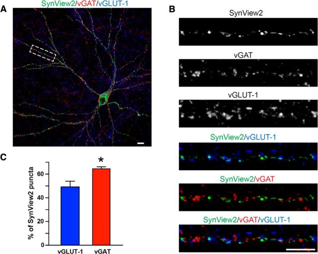
Figure 9.
Analysis of SynView2-positive synapses in cultured neurons. A, Representative image of a transfected hippocampal neuron expressing NL2-GFP11S2 that is surrounded by neurons expressing Nrx1β-GFP1–10S2. Punctate SynView1 GFP fluorescence (green) was combined with immunofluorescence staining for vGAT (red) and vGLUT-1 (blue). Scale bar, 10 μm. B, High-magnification images of the boxed dendritic segment in A showing individual and composite images of the different stainings. Scale bar, 10 μm. C, Quantitative analysis of the percentage of SynView2-positive puncta that are adjacent to vGLUT1- or vGAT-positive puncta. Data represent means ± SEM; n = 3 independent experiments; *p < 0.05. Data show representative experiments independently repeated at least 3 times.

