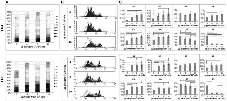
Figure 2.
The proliferative ability of in vitro stimulated PBLs is reduced by exo-hASCs. The PBLs were cultured either alone or co-cultured with different batches of exo-hASCs (n = 8) at different concentrations (4, 8, and 16 μg of exosomes per million of PBLs). At day six, PBLs were collected and T-lymphocytes subsets were stained with anti-CD3, anti-CD4, and anti-CD8. Fluorescence profiles of CFSE-labeled cells allowed us to identify eight divisions. A detailed representation of CD4+ T cells and CD8+ T cells showing the percentage of the total population in each cell division cycle (indicated as #) is provided (A), as well as a representative histogram (B). The statistical comparison of lymphocyte subsets at different cell division cycles is also provided (C). Horizontal bars represent statistically significant differences between the groups (significant at p ≤ 0.05).

