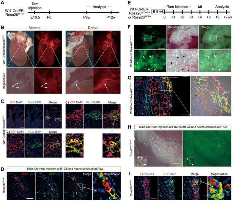
Figure 1.
Epicardial cells differentiate into adipocytes during cardiac development and injury. (A) Strategy of inducible labeling of epicardial cells. Tamoxifen was administered at E10.5 to Wt1-CreER; Rosa26RFP/+ embryos, and adult hearts were collected for analysis. (B) Whole-mount view of fat tissue in atrioventricular groove (arrows). (C) Sections stained for lineage marker RFP, adipocyte marker PLIN, and nuclei marker DAPI. C1 and C2 show that a subset of adipocytes in the right and left atrioventricular grooves were derived from epicardial cells. Images are representative of results from four individual hearts. (D) Co-immunostaining of PLIN and GFP in P8w Rosa26mTmG/+ heart treated with Msln-Cre adenovirus at E12.5. (E) Strategy for lineage tracing of adult epicardial cells after MI. Adult mice were treated with tamoxifen before MI. (F) Whole-mount picture of adult labeled heart following MI. Arrowheads indicate epicardium-derived adipocytes within fat tissue on heart surface. (G) Immunostaining of post-MI heart sections shows that a subset of PLIN+ adipocytes co-express the GFP lineage marker. (H) Msln-Cre adenovirus was injected into the adult heart. After MI, whole-mount images show lineage-traced GFP+ cells within fat tissue on the heart surface. (I) Immunostaining of PLIN and GFP in Msln-Cre adenovirus-treated Rosa26mTmG/+ hearts after MI. Red bar = 0.5 mm; yellow bar = 200 μm; white bar = 100 μm.

