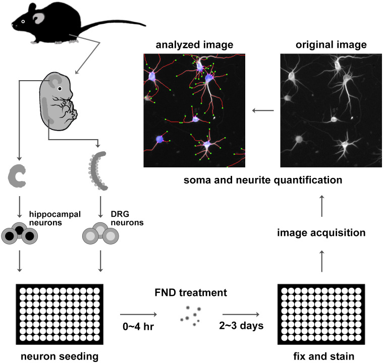
Figure 1. Schematic diagram of dissociated primary neurons preparation, fluorescent nanodiamond treatment, and imaging procedure.
Primary neurons from mouse hippocampi and the dorsal root ganglia were isolated from embryonic mice, dissociated with protease, and seeded into 96-well plates. Dissociated neurons were treated with FNDs for 2 ~ 3 days, fixed, and immunofluorescence stained with antibody against neuron-specific β-III-tubulin. Automated image acquisition and analysis were performed on stained neurons.

