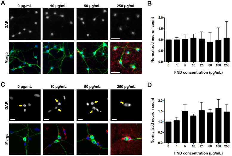
Figure 2. Fluorescent nanodiamonds did not cause cytotoxicity in dissociated hippocampal or dorsal root ganglion neuron cultures.
(A) Images of dissociated hippocampal neurons treated with various concentrations of FNDs 4 hours after seeding and incubated for 3 days in vitro. Images on the top row show DAPI-stained nuclei, merged images on the bottom row show FND (red), neuron-specific β-III-tubulin (green), and DAPI-stained nuclei (blue). Scale bars represent 50 μm. (B) Quantification of neuron number per image field of FND treated hippocampal neurons. (C) Images of dissociated DRG neurons treated with various concentrations of FNDs after seeding and incubated for 2 days in vitro. Arrows point to the nuclei of DRG neurons. Scale bars represent 20 μm. (D) Quantification of neuron number per image field of FND treated DRG neurons. All quantification data were obtained from 3 independent repeats and normalized to the group without FND. No statistically significant difference between control group and FND-treated groups can be detected. p > 0.05, by one-way ANOVA. Both bar graphs are expressed as mean ± SEM.

